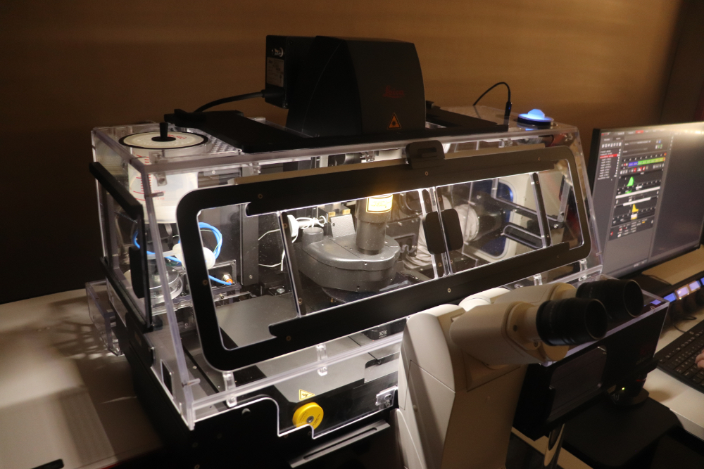
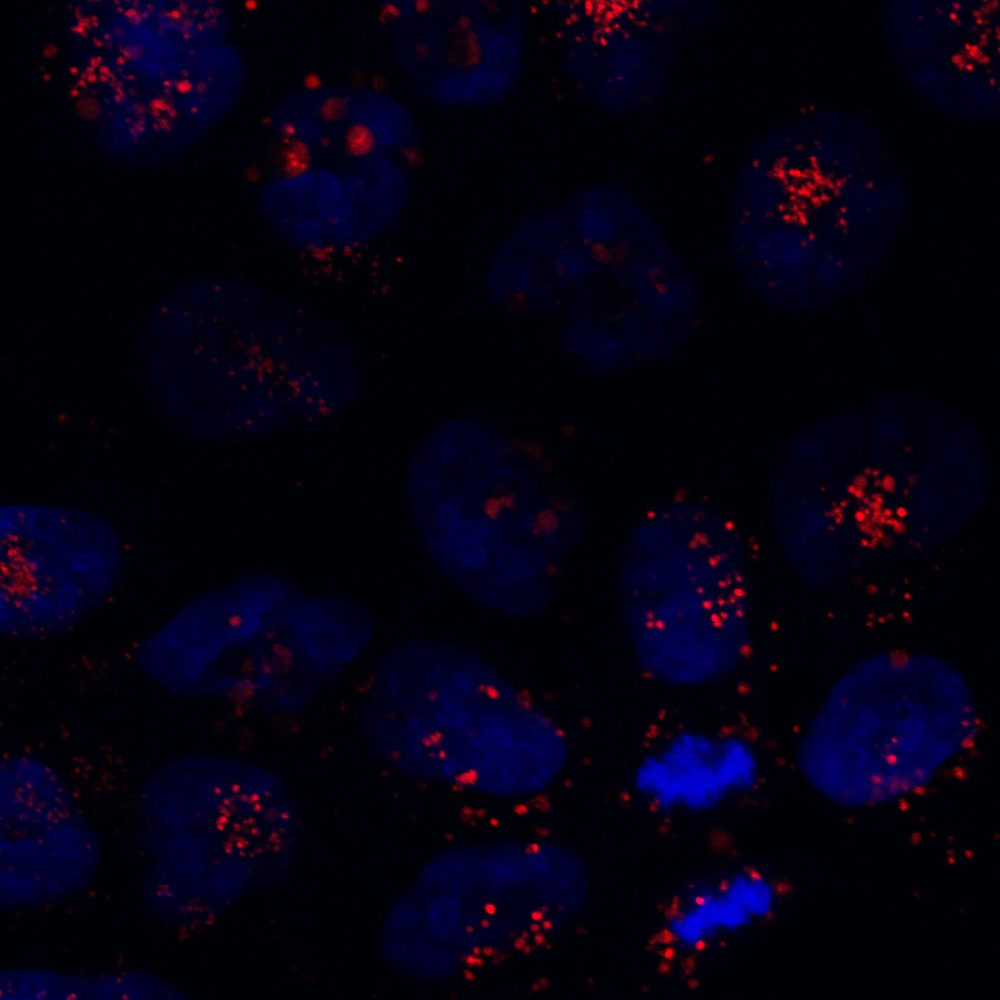
Description
MICROSCOPE:
Leica DMI8;
Fully motorized inverted microscope;
Universal adapter for slides and 35” Petri dishes;
OKO lab Cage incubation system including CO2 Unit with active humidity controller;
OBJECTIVES (all with special APOCROMATIC PLAN correction for confocal techniques):
10x NA 0.40 dry;
20x NA 0.75 dry;
40x NA 1.30 oil;
63x NA 1.40 oil;
CONFOCAL:
White Light Laser Stellaris 8 (440-790nm);
UV (diode 405 nm/50mW);
Acquisition of multidimensional series: x, y, z, t, λ and their combinations;
High resolution scanning system (maximum resolution: 8,192 x 8,192 pixels; maximum line frequency: 3,600 Hz; minimum line frequency: 1 Hm; image frequency at 512 x 512: 9 images / sec., full field; image frequency at 512 x 16: 84 images / sec., full field; “beam park” reading mode; optical zoom: 0.75x ... 48x; field diameter: 22 mm);
Three spectral detectors, controlled independently in terms of gain, offset, … ;
Automatic Beam Splitter able to combine more than 4 or 5 wavelengths;
Super-resolution spectral detection system (lateral resolution better than 120 nm; axial resolution better than 200 nm; more than 20 images by second with super resolution);
FLIM (Fluorescence Lifetime Imaging Microscopy):
Two cooled FLIM spectral detectors;
Acquisition of FLIM images in different dimensions, time lapse, 3D, lambda, mosaics, etc.;
FLIM / FRET module;
Measurements with deadtime full system 1.5 ns ;
2D graphic display of the life times that allow the selection of the life times of interest in the graphic itself by defining regions;
Separation of fluorochromes by FLIM, based on life-time calculations;
APPLICATIONS:
Confocal imaging;
Live cell imaging;
3D imaging;
FLIM;
FRET (Förster Resonance Energy Transfer);
FRAP (Fluorescence Recovery After Photobleaching).


